A. process sensory information from internal organs, from environments
B. decide what to do about it.
C. Do something. Move skeletal muscles or internal organs, change biochemical reactions in internal organs
D. assume you know parts of nerve cells.
II. Use of disk. Forebrain not brain stem or spinal cord. Can see structures in various orientations.
A. choose sections, then click on building, then on system you are interested in.
B. click on key to see what colors mean. You may not see a struc in all orientations.
C. use index to find a strucutre.
D. can see how brain fits into head by clicking on orientation.
E. assume you know orientation terms, if not see pg 36.
III. Show spinal cord in brain and 3 different views of brain.
IV. Spinal Cord
A. continuous with brain stem
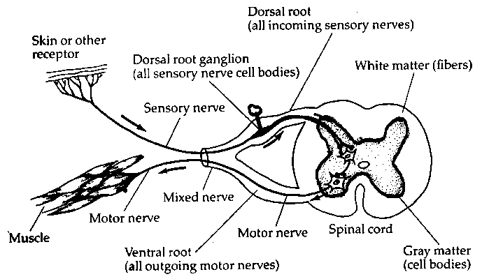
B. Parts of the spinal cord provides clear information of how info gets into and out of brain.
1. dorsal horn
2. ventral horn
3. ventral root
4. dorsal root.
5. dorsal root ganglion
6. sensory nerve
7. motor nerve
8. mixed nerve
see diagram:

C. Be sure you can draw how dorsal root axons enter cord and synapse with dorsal horn neurons.
D. Be sure you understand how ventral root axons leave the cord.
V. Peripheral nervous system
A. sensory nerves are afferent
B. motor nerve are efferent
C. mixed nerve have both afferent and efferent fibers.
VI. Motor: skeletal
A. Motor cortex to spinal cord
B. to subcortical nuclei
C. Basal ganglia
D. cerebellum
VII. Motor: smooth. Internal organs are tightly controlled by the brain. Stress parallel with skeletal motor system.
A. Autonomic nervous system (you need not know all the ganglia and how they connect to internal organs but you need to understand the general plan of the autonomic nervous system.) It consists of 2 parts, the sympathetic and the parasympathetic nervous systems.
B. The sympathetic nervous system is active during flight or fright, when the organism is aroused.
1. sympathetic nervous system originates in the thoracic and lumbar parts of the spinal cord
2. the neurons that provide inputs to the sympathetic ganglia are located in the lateral parts of the thoracic and lumbar spinal cord. These are preganglionic neurons
3. the motoneurons for the sympathetic nervous system are in the sympathetic ganglia which form a chain adjacent to the thoracic and lumbar parts of the cord. These motor neurons secrete norepinephrine. They innervate internal organs.
C. parasympathetic nervous system - active during relaxation
1. originates in the cranial and sacral parts of the spinal cord
2. preganglionic neurons provide input to the parasympathetic ganglia. located in the lateral parts of the cranial and sacral parts of the spinal cord.
3. motoneurons are in the parasympathetic ganglia which are clusters of neurons near the organs that they innervate.
D. Comparison of sympathetic and parasympathetic function.
1. all organs receive both types of innervation.
2. depending on the function of an organ, it will be inhibited by one system and excited by the other.
3. For example: heart must accelerate when the organism is aroused and can beat more slowly during periods of relaxation. Therefore it is excited by the sympathetic system and inhibited by the parasympathetic system.
4. For example: stomach must stop digesting food when the organism is aroused to escape from an enemy because the muscles need the blood supply. It can go back to digesting during periods of relaxation. Therefore it is excited by the parasympathetic system and inhibited by the sympathetic system
VIII. The following items may show up on the exams even though we did not discuss them in class. You should know the location and function of each structure:
A. spinal cord
B. inputs and outputs of brain: cranial nerves.
1. can be sensory or motor or both
2. can enter or leave brain at medulla, pons, or junction of pons and midbrain
C. medulla: vegetative function like heart rate, respiration, provide input to autonomic ns
D. midbrain:
1. tectum
a. superior colliculus-vision
b. inferior colliculus-hearing
2. tegmentum
a. red nucleus and substantia nigra: important in movement.
E. pons: some nuclei important in sleep and arousal, some in transferring motor information between motor cortex and cerebellum.
F. diencephalon:
1. thalamus-know how to locate the thalamus in the brain. sensory processing and other less well understood functions.
2. hypothalamus
3. pituitary (not actually part of diencephalon)
G. cerebellum
H. Cerebral cortex
1. occipital lobe
2. temporal lobe
3. parietal lobe
4. frontal lobe
5. mapping of sensory space onto somatosensory, visual, auditory cortex.
6. mapping of muscles onto motor cortex.
I. Structures in cerebrum
1. basal ganglion
2. limbic system and hippocampus
3. corpus callosum
J. Fluid supply to brain-ventricles